


A cell needs to generate forces to control the angle of division axis by employing molecular force generators. To date, the microtubule motor protein dynein is the only known force generator that pulls microtubules to orient the mitotic spindle. However, our knowledge of cell division orientation is limited due to the lack of multicellular studies at single cell resolution. In this study, Sugioka and Bowerman studied multiple cell types of C. elegan embryos and found the cell division orientation mechanisms independent of the microtubule-dynein pathway. Analysis of multicellular division is challenging; an individual cell is surrounded by different physical and chemical cues, limiting our ability to identify the causal relationships between upstream cues and division axis. To overcome this predicament, Sugioka and Bowerman reconstituted cell contact patterns using single cells isolated from living-embryos and adhesive polystyrene beads. They identified three contact-dependent cues that specify distinct cell division axes: single cell contact, asymmetry of contact sizes, and the Wnt signal.
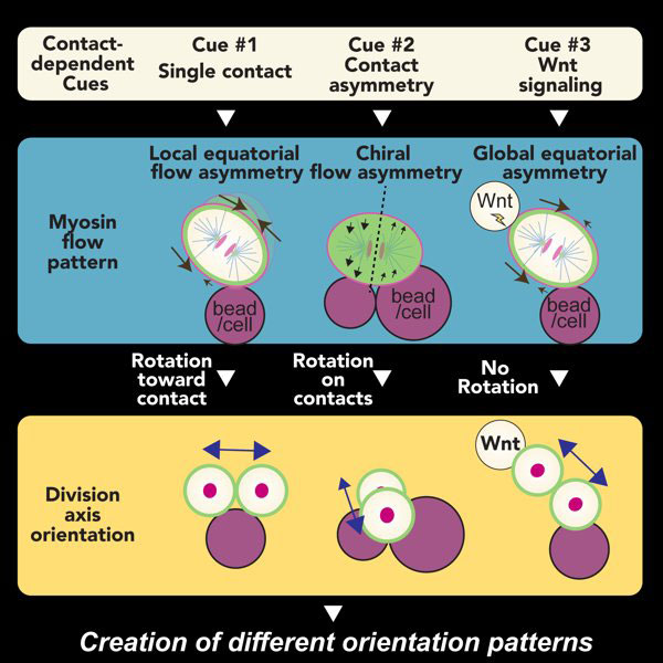
Figure: Schematics of contact cue-dependent cell division orientation. Three contact cues (top) control different myosin flow patterns (middle; black arrows) to specify distinct cell division axes (bottom; navy blue arrows).
The three contact-dependent cues generate the distinct patterns of cell surface myosin flow; myosin flow, or cortical flow, is a collective flow of the actin-myosin network at the cell surface. However, it has not been experimentally shown that myosin flow can generate forces to specify division axis. Sugioka coated the outer cell surface with micro-particles and tracked their movements relative to myosin flows, and showed that myosin flow could trigger cell surface movements by generating forces.
Interestingly, Sugioka also found that mouse 2-cell stage embryonic divisions are also regulated by contact-dependent cues and myosin activity. These results suggest that contact-dependent cell division orientation mechanisms are evolutionarily conserved in animal development. A big unanswered question is “how myosin interprets different contact patterns?” Future study should uncover the mechanism of such a cellular decision-making process that orchestrates division axis diversity.
Reference
Combinatorial Contact Cues Specify Cell Division Orientation by Directing Cortical Myosin Flows. Sugioka, K., and Bowerman, B. (2018). Developmental Cell 46, 257-270.e5. (Cover article).
Preview article (Developmental Cell)