


In multicellular organisms, forms seen in the adult find their origins during development, where a blueprint of gene expression foreshadows the final morphology. For instance, the spatial expression of developmental genes in the wing precursors of butterflies during metamorphosis prefigure their diverse colour patterns. In turn, the spatial distribution of gene products (transcripts), results from the activity of DNA segments flanking these genes and known as gene switches or transcriptional enhancers. It has long been known that these regulatory elements are bound by particular proteins –transcription factors– that control them and modulate the levels of gene transcription across a tissue, thereby producing a gene expression pattern. While transcription factors recognise specific stretches of DNA on an enhancer –binding sites–, how a particular organisation of binding sites on an enhancer results in a given expression pattern is unclear.
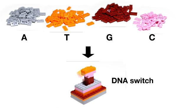
Figure: How is a transcriptional enhancer built? In this study, using mutations in a simple regulatory element, and monitoring its resulting activity quantitatively, we deconstructed its regulatory logic to its essential components, the nucleotides. This is another way to ask how the 4 letters that compose DNA can be arranged to build such a sophisticated biological object, much like simple Lego pieces can be used to build complex devices. Copyright: N. Gompel.
Supported by an HFSP Program Grant, we have joined forces to decipher the regulatory logic of a model enhancer, combining genetics, molecular biology and the quantitative analysis of images reflecting enhancer activity. Our model enhancer, conveniently called spot enhancer, drives the expression of a pigmentation gene in a spot pattern at the wing tip in flies (Drosophila). We visualised the activity of the spot enhancer in developing wings by placing a gene encoding a fluorescent protein under its control. Hence, the distribution of fluorescence, at different levels across a fly wing reveals the spatial activity of the spot enhancer. To decipher how this enhancer is built, we introduced mutations along its sequence, or challenged its structure by shuffling blocks of sequence. This is roughly equivalent to taking pieces out of a motor, or tinkering with them and reorganising them to understand the causal relationships between each piece and the motor in motion.
A fairly unique and novel aspect of our approach is that we did not simply compare each mutant to the wild-type spot, in a pair-wise manner. Instead, we devised a method to warp all wing images onto a reference wing, such that the activity pattern of all mutants became comparable in the same framework. This was key to understanding the contribution of each mutated site, no matter how subtle, to the whole activity. Two main messages emerged from our work. First, the density of regulatory information was far greater than generally assumed in the literature on transcriptional enhancers. Every site along the spot enhancer sequence, which we scanned, contributed to the whole activity. Second, the simple pattern driven by this enhancer, a two-dimensional wing spot, resulted from an unexpectedly complex molecular construction, with multiple tiers of transcription factors interacting, repressing each other, to carve the elementary spatial pattern.
There is more to unravel to understand the relationship between the different partners in the multi layered regulation of this model enhancer. Our recently published results represent a first step, and more importantly a framework to refine our dissection. Funding from HFSP has been instrumental in building this framework, bringing our complementary expertise in molecular biology, structural analysis of the DNA sequence, and advanced image processing together.