


PET imaging offers excellent translational possibilities due to its combination of sensitivity and quantitative accuracy. It provides very useful information about tissue and organ function and status, and the type of physiological information provided depends on the specific imaging agent used. The process begins with the injection of a radioactive compound called radiotracer (at a harmless low dose) followed by the detection of the positron-mediated gamma radiation emitted. The positrons travel a certain distance in tissue until they annihilate into the two gamma photons detected by the scanner. This distance or “positron range” (PR) is one of the main sources of PET imaging blurring, but its effect could be remedied to a large extent if accurate estimates of this phenomenon are considered during the image reconstruction process.
A.  B.
B. 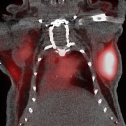
C. 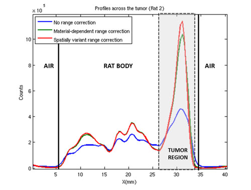
Figure: PET Image Quality Improvement Using Tissue-Dependent, Spatially-Variant Positron Range Correction. Images show a coronal slice of the PET/CT image of a mouse thorax with xenografted neuroendocrine tumors (NET). The CT image is depicted in black and white while the white and red is the resulting PET image for the 68Ga-DOTATOC radiotracer that binds to the somatostatin receptor expressed by the NET (contrast is enhanced for better visualization). (A) Non-PR corrected reconstruction produces a blurrier image with less defined contours plus a background haze that limits the accurate delineation of the tumor (hot area under the right foreleg); (B) PR-corrected image where the haze has disappeared and the tumor can be better delineated; (C) Horizontal profile across the mouse thorax depicting the PET image intensity showing how the PR-correction improves the uptake quantification on every tissue.
Previously proposed methods to remove the PR effect in PET images have proved to be only partially effective because they ignored the boundaries between tissues of different density. The solution proposed in this work models PR effects in the image reconstruction algorithm as 3D anisotropic (spatially-variant) blurring kernels based on the tissue density-dependent radial profile of the annihilation points. This tissue-dependency is extracted from the CT image that is acquired together with the PET. If cleverly implemented, this approach has a small computational cost, it improves significantly the quality of the reconstructed images, rendering material-dependent PR corrected images, and removes edge artifacts frequently seen in clinical and preclinical images.
This advanced algorithm produces better quantification of the lesions of interest, giving activity values closer to the theoretical ones. Regarding mice studies, the quantification of tumor activities improves significantly because it allows a more precise delineation of the different tissue boundaries, among other reasons.
The computation of this algorithm adds a manageable overhead to the overall reconstruction time. Most of this overhead is due to the kernel pre-calculation from the CT image, but this needs to be done only once per acquisition, even for dynamical multiframe images. This implementation allows for straightforward parallelization and it can be implemented to exploit multi-core and multi-CPU execution, or GPUs with significant speed-ups that yield total reconstruction times of a few minutes, even for the largest positron range case.
The proposed method implements an accurate and efficient spatially-variant PR correction that provides accurate, artifact-free reconstructed images. This is especially valuable in situations where the activity is located close to tissue-boundaries or in heterogeneous media. It improves significantly the quality of the reconstructed images for medium and large PR radionuclides. Although this improvement on image quality is particularly relevant when working in high-resolution imaging with small-animal models, it can also improve quantification accuracy in clinical PET imaging, even in the presence of magnetic fields (PET/MRI systems).
Reference
Tissue-dependent and spatially-variant positron range correction in 3D PET. Jacobo Cal-Gonzalez, Mailyn Perez-Liva, Joaquín Lopez-Herraiz, Juan José Vaquero, Manuel Desco, José Udias. IEEE Transactions on Medical Imaging 62, 2015, doi = 10.1109/TMI.2015.2436711.