


During development embryonic cells need to achieve two major tasks. First, they need to differentiate into specific cell types. Second, they need to organise in space to shape tissues and organs with precise forms and functions. A striking example of this is the development of the heart. To achieve the formation of the heart, embryonic cells must differentiate into cardiac cells and first establish a longitudinal “primitive” tube, called the heart tube, which pumps early on to supply oxygen and nutrients to the developing embryo. The primitive heart tube then further develops into the more mature four-chambered heart during later steps of embryonic development.
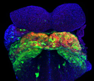
Figure: Mouse embryo showing heart-forming cells labeled with fluorescence for two-photon microscopy. Green-only cells are SHF cells and cells that are both green and red are FHF cells. Credit K. Ivanovitch.
How the heart tube forms and how its formation relates to the timing of cardiac differentiation is largely unknown. To address this question, Ivanovitch et al. live-imaged for the first time the embryonic cells that differentiate into cardiac cells and form the primitive heart tube in intact mouse embryos. To achieve this, they cultured mouse embryos ex vivo and used advanced microscopy and cell tracking techniques. They discovered unexpected temporal regulation of cardiac differentiation and its coordination with heart tube formation. During an initial phase, embryonic cells differentiate rapidly into cardiac cells, while limited cell movements take place. During a second phase, no differentiation events are detected, while extensive cellular migration and rearrangement results in the formation of a heart tube, still open dorsally at this point. During a third phase, cardiac differentiation resumes and the closure of the heart tube is completed. These results therefore reveal an uncoupling between morphogenesis and differentiation during HT formation.
Much of what is known about heart development in mammals is based on the analysis of fixed embryos. However, in their new research, Ivanovitch et al. recorded movies of the developing heart in mouse for the first time and important observations on the way cells differentiate and rearrange to form the heart tube over time have been revealed. The methods used in this study bear an enormous potential. They will help reveal why abnormal heart develops in certain cases, and they can be applied to the study of other organogenesis processes in the future.
Video: The morphogenesis of the heart tube during the initiation of the second phase. FHF cells are shown in green and SHF cells are highlighted in red. Credit K. Ivanovitch.
Reference
Live imaging of heart tube development in mouse reveals alternating phases of cardiac differentiation and morphogenesis. Kenzo Ivanovitch, Susana Temiño Valbuena, Miguel Torres. eLIFE, Research Article Dec 5, 2017, https://doi.org/10.7554/eLife.30668.