
Mapping intracellular phase space using photo-activated protein droplets

Eukaryotic cells compartmentalize their contents into diverse specialized organelles, many of which can maintain their distinctive composition of mobile biomolecules even in the absence of a physical barrier such as a lipid membrane. In recent years, various membrane-free organelles were found to display liquid like properties, suggesting that they were the product of a liquid-liquid phase separation process, in which concentrated aqueous condensates of weakly interacting biomolecules coexist with a surrounding dilute aqueous phase. However, in the absence of quantitative tools for controlling and studying phase transitions in live cells, the underlying biophysical principles are still largely unknown, particularly for phase separating proteins of low-abundance. Moreover, whether phase separation can be truly maintained under the out-of-equilibrium intracellular environment remains unclear.

In a recent study we have introduced a biomimetic system for reversibly driving intracellular phase separation in a spatiotemporally controlled manner using photo-induced oligomerization of weakly interacting proteins. We utilized the quantitative nature of this platform to map, for the first time, full intracellular phase diagrams, which deterministically dictate whether and how phase separation occurs and quantitatively describes how specific mutations and post-translational modification modulate the transition. Moreover, we show that while intracellular protein concentration may be insufficient for global phase separation, locally tuning the conjugation number of phase-separating proteins to slowly diffuse scaffolds can move the cell into a different region of the phase diagram, resulting in localized phase separation. We suggest that this diffusive capture mechanism liberates the cell from the constraints of global protein abundance and is likely exploited to pattern condensates associated with diverse biological processes.

Bracha2019a


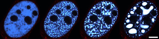
Figure: Nucleus of a representative U2OS type cell (human bone cancer) undergoing phase separation. The transition from a uniform single-phase state (left) into a two-phase state, in which condensates of concentrated proteins (white blobs) coexist with their dilute surrounding, is initiated by blue light activation and lasts for two minutes. Scale bar is 10 µm.

Reference

Mapping Local and Global Liquid Phase Behavior in Living Cells Using Photo-Oligomerizable Seeds. Dan Bracha, Mackenzie T. Walls, Ming-Tzo Wei, Lian Zhu, Martin Kurian, Jose´ L. Avalos, Jared E. Toettcher, and Clifford P. Brangwynne. (2019) Cell, 175, 6, 1467-1480.

Press release from Princeton University

Preview paper in cell

Highlight paper in Nature methods

Media contacts

Guntram Bauer
Director of Science Policy and Communications

Liliana Gonçalves
Science and Communications Officer

Rachael Bishop
Science Writer and Editor

Click here to show mail address


Reference

Mapping Local and Global Liquid Phase Behavior in Living Cells Using Photo-Oligomerizable Seeds. Dan Bracha, Mackenzie T. Walls, Ming-Tzo Wei, Lian Zhu, Martin Kurian, Jose´ L. Avalos, Jared E. Toettcher, and Clifford P. Brangwynne. (2019) Cell, 175, 6, 1467-1480.

Press release from Princeton University

Preview paper in cell

Highlight paper in Nature methods