


Resolution in optical microscopy has been fundamentally limited by the wave nature of light to about 200 nm. This changed with the advent of super-resolution microscopy methods, improving the resolution by one order of magnitude. Recently, the group of Nobel laureate Stefan Hell developed a revolutionary new super-resolution microscopy method called MINFLUX [1], that now allows us to reach a resolution on the size of a small molecule, revealing unprecedented detail when imaging cellular structures. Now, Hell and coworkers extended this technology to 3D and dual-color and to extended fields of view.
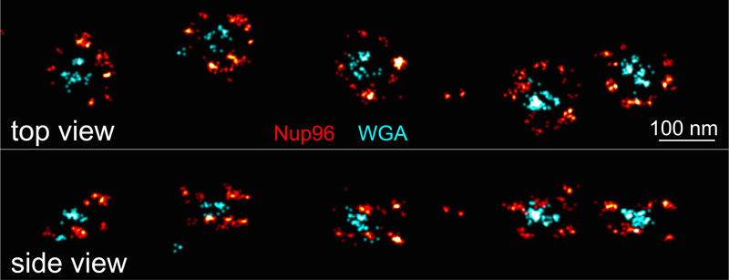
MINFLUX image of the nuclear pore complex. Three-dimensional dual-color MINFLUX data of the nuclear pore scaffold protein Nup96 and the label of the central channel WGA (top and side views) reveal the precise architecture of this complex protein machine with unprecedented optical resolution.
To demonstrate the power of these new developments, a precise cellular structure was needed, thus the Hell group teamed up with the group of HFSP Young Investigator Jonas Ries who had established super-resolution reference standards as part of his HFSP grant [2]. These reference structures consisted of nuclear pore complexes, the channels that regulate transport between the nucleus and the cytoplasm of cells, which the Ries group had labeled with super resolution-compatible fluorophores using the CRISPR/Cas9 technology. The new MINFLUX microscope could reveal the structure of these complex protein machines with unprecedented optical resolution. As a major breakthrough, this was even possible in living cells, paving the way for imaging molecular machines during their action.
This new technology has the potential to revolutionize how we can study the building blocks of life inside cells during their function and provides numerous applications in cell and structural biology.