


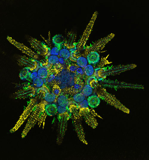
Sea urchins do not possess a central neural control center or brain. Their behavioral repertoire, however, is rather complex. This is especially true for the urchin's reaction to light. Not only can they detect looming visual stimuli from any direction and accurately point their spines towards them, but they are also able to resolve objects and move straight in their direction. What adds to the mystery of these behavioral capabilities is the fact that sea urchins are eye-less and instead feature dermal photoreceptors spread over their entire spherical body.
The discovery that sea-urchins may use photoreceptor cells distributed around their whole body for vision was made in 2011 when a team led by Maria Ina Arnone, working at Stazione Zoologica Anton Dohrn in Naples, Italy, characterized for the first time photoreceptor cells in a sea urchin (www.pnas.org/content/108/20/8367). The results of the 2011 paper led to the speculation that sea urchins must have a sort of decentralized visual system, which may work like a giant compound eye, that allows them to detect light stimuli. The question arose: how can the sea urchin, which possesses only a decentralized nervous system distributed along 5 principal nerves connected by a nerve ring around their mouth, integrate external light stimuli and respond to them?
To answer this question, an integral approach is required that combines molecular, morphological, electrophysiological, behavioral and computational studies. This is exactly what an international team of scientists led by Dr. Arnone plans to do. The team includes Prof. Dan-Eric Nilsson of Lund University in Sweden, an expert in animal vision; ecologist and morphologist Carsten Lüter of the Museum für Naturkunde in Berlin, Germany and Giancarlo La Camera, a computational neuroscienstist of the State University of New York at Stony Brook, USA. The four scientists will work together to tackle this challenge largely thanks to an HFSP research grant awarded in 2019.
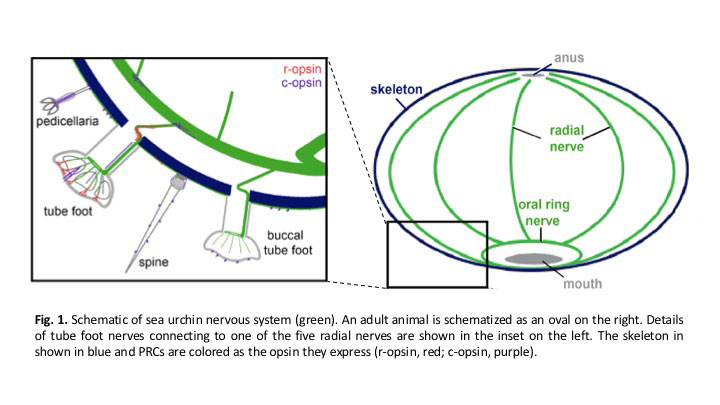
The ultimate goal of the project is to obtain a computational model of the decentralized “spherical” vision of the sea urchin from its molecular, morphological and physiological makeup. To put this challenge into perspective, one would expect an integrated neural control center such as a central nervous system to make sense of the information coming from so many light sensitive cells distributed around the whole body. However, there aren’t any in the sea urchin. Instead, a de-centralized arrangement of five radial nerves connected to a nerve ring around the animal’s mouth is the structure that does the job.
To understand this system, one needs to understand a number of related mechanisms: 1) what types of neurons are present in the radial and oral ring nerve, how they are connected, and how they exchange information; 2) how are light stimuli processed by the photoreceptor cells; 3) how is the information processed by the photoreceptors sent to the neurons in the radial nerves. These are basic molecular, morphological and electrophysiological properties that are necessary to establish in order to make a working model of vision. Behavioral assays will also be necessary to establish the resolution of the visual system – the dimension of detectable visual stimuli and the extent the animal will move towards or away from them. This information has high ethological value, as it establishes what the animal can and cannot do based on visual stimuli in its environment.
Next, to obtain a more refined model, the specific mechanisms of information exchange between stimuli, photoceptors, neurons and motor output must be understood. Perhaps surprisingly, little is known about the elements essential to uncover these mechanisms. The 5 radial nerves are connected to an oval ring, but the anatomical details are unknown. Neural cell types are also unknown; neurons are broadly divided into excitatory and inhibitory, according to the type of action they exert on their target neurons, and more refined subdivisions exist. The team will have to establish what type of neural cells make up the radial and the oral nerve together. They will also have to establish their pattern of connectivity, i.e., which cell types are preferably linked together, and in which direction. Do neurons receive only direct input from earlier stages, as in a feed-forward network architecture, or is there evidence of “recurrent” feedback from neurons in the same nerve structure, as it is known to be the case in single layers of the mammalian cortex?
Another mechanism that will have to be determined is whether sea urchin neurons communicate preferably via chemical or electrical routes. Mammalian neurons can exchange electrical information via chemical synapses (the predominant method) or via electrical contact points called ‘gap junctions’. Which one is the case for the sea urchin?
These considerations show that a large amount of experimental information will have to be gathered in order to construct a coherent understanding of decentralized vision in this fascinating creature. This is the reason why the team comprises expertise at several levels spanning molecular to behavioral. The team is confident that, in addition to aiding a computational model of decentralized vision, their work will provide the wider community with a great deal of information on yet unknown molecular, morphological and behavioral aspects of echinoderms’ biology.
Decentralized vision itself, using the sea urchin as a model organism, can prove useful beyond the biological realm, as it may lead to applications in the field of bio-mimetics. Potential biomimetic applications include robotic miniaturization, smart probes, and intelligent materials where dispersed light detectors control the properties of the material. It’s too early to say how much we will learn from this venture, but the road is full of exciting possibilities.