


Cheap and fast tools for gene sequencing, protein expression and analysis are routinely used for high-throughput genomics (1,2). However, protein structure determination cannot presently be performed at the same fast pace, due to experimental difficulties and longer timescales (e.g. for crystallization): a fact that is slowing down the discovery of new protein features as well as their engineering (3). These difficulties heavily affect the field of photobiology, where the number of known light-responsive protein structures is limited. For instance, the crystal structure of bovine rhodopsin, the retinal dim-light visual photoreceptor protein, is the only structure available for studying monochromatic and color vision in vertebrates. This impairs our understanding of the basic relationship between protein sequence and photoreceptor function, which requires a comparison of diverse photoreceptors. This problem becomes paramount in evolutionary studies where one wishes to compare entire collections of ancestral sequences revealing how adaptive specializations may have occurred in nature.
The above issues can, in principle, be overcome by constructing atomistic computer models of the set of proteins of interest, accurate enough to allow for in silico predictions of function, screening different protein properties and the discovery of novel design. When focusing on ancestral proteins, this requires the integration of different technologies such as computational molecular evolution, comparative modeling and quantum chemistry. Our collaborative HFSP funded research goal was to use a multi-disciplinary approach to studying the large family of light-responsive proteins: rhodopsins. The rhodopsin family is an excellent target for these studies since they display wide ranges of spectral and reactivity changes mediated by the same covalently bound, vitamin A-derived moiety called the chromophore. The protein sequence must control two fundamental steps in rhodopsin activation: absorption of light energy and its utilization for the isomerization of a specific double bond of the chromophore.
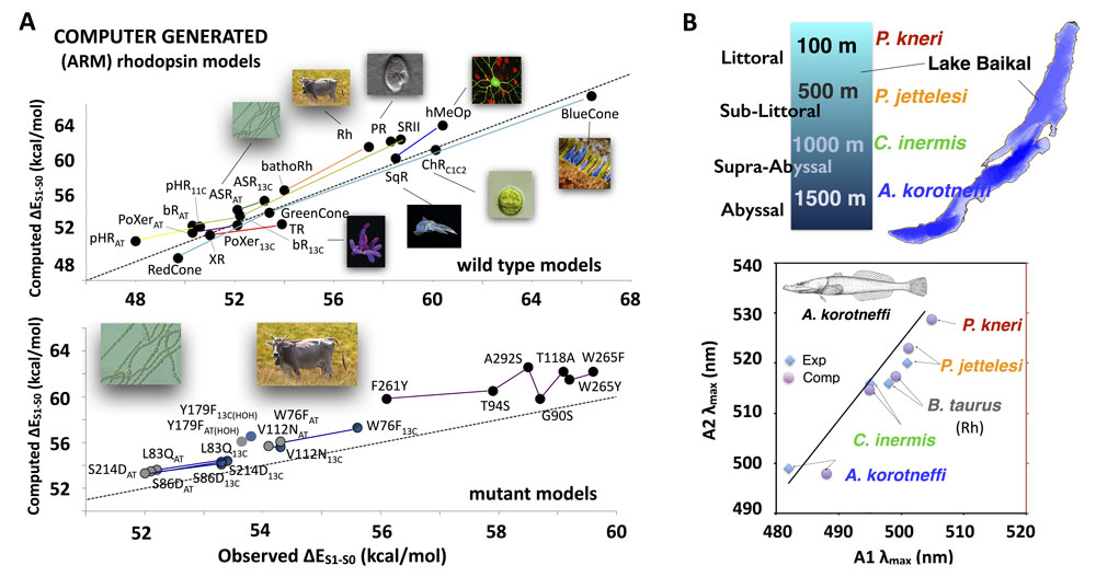
The research started by designing a computer program capable of automatically generating hybrid quantum mechanics/molecular mechanics rhodopsin models of the required accuracy. To be useful, such models must be produced using a fast (several hours/model), well defined and replicable protocol with known error bars. Full sets of rhodopsin models would then be generated on a parallel computer exactly on the same time-scale. The first version of such a computer program, called ARM (Automatic Rhodopsin Model), has been recently reported (4). The benchmarking studies (see Figure A) validated our ARM models by reproducing the trend in color sensitivity (i.e. the excitation energies ΔES1−S0) for rhodopsins of a wide range of different organisms.
(click on image to enlarge)
Figure: Benchmarking and application of the ARM program. (A) Observed vs computed values for vertical excitation energies (ΔES1−S0=h∙c/λmax) of wild-type rhodopsins. Full colored lines connect related rhodopsins. Bovine rod rhodopsin (orange), human cone rhodopsin (light blue), invertebrate and nonvisual rhodopsins (blue), the eubacterial sensory rhodopsin from Anabaena (green), Prvularcula (brown) and archaea rhodopsins: bacteriorhodopsin and sensory rhodopsin II (dark yellow), xanthorhodopsin and Thermophilus rhodopsin (red), pharaonis sensory rhodopsin (light yellow). Channelrhodopsin ChRC1C2 is the only rhodopsin originating from microbial eukaryotic rhodopsins. The eubacterial proteorhodopsin (PR) is also found in eukaryotes (Dinoflagellates). Comparison between computed and observed ΔES1−S0 values of a set of Anabaena (left part of the diagram) and bovine rhodopsin (right part of the diagram) mutants. (B) Top. A recently reported application focusing on the evolution of the visual pigment of different Cottoid fish species thriving at different depths (i.e. illumination levels) in lake Baikal. Bottom. λmax of Baikal cottoid fish rhodopsins reconstituted with the A1 (wild type) and A2 chromophores. Experimental and computed λmax of the A1 and A2 models respectively. The straight line indicates the linear relationship of 18 identical-opsin pairs previously reported by Dartnall and Lythgoe (5).
Our ARM technology has allowed us to complete the first molecular-level evolutionary study of dim-light vision. The study (see Figure B) focused on the fauna of Lake Baikal: one of the most ancient and deepest lakes in the world. Its unique ecology has resulted in the colonization at a diversity of depth habitats by a unique fauna that includes a group of closely related teleost fish of the sub-order Cottoidei. This relatively recent radiation of cottoid fishes shows a gradual blue-shift in the wavelength of the absorption maximum of their visual pigments with increasing habitat depth (6). We combined comparative modeling and ARM calculations with experimental in vitro measurements of the Lake Baikal rhodopsins to investigate such dim-light adaptation (7). The calculations, which were also able to reproduce the trend of observed absorption maxima shifts by changing the chromophore (A1 to A2), reveal a previously hypothesized (Barlow) relationship (8) between the absorption maxima and the thermal isomerization rate suggesting a link between the observed blue-shift in the spectral sensitivity and a decreased thermal activation of the rhodopsin. Based on the results of a point-charge analysis of the electrostatic effects of non-conserved and conserved amino acids, we proposed that natural variation at these sites modulates both the thermal noise and spectral shifting in Baikal cottoid visual pigments resulting in adaptations that enable vision in deep and dim water light environments.
References
1. High-Throughput Protein Production (HTPP): A Review of Enabling Technologies to Expedite Protein Production. Doyle, S., Koehn, J. & Hunt, I. in High Throughput Protein Expression and Purification 1-18 (Humana Press, Totowa, NJ, 2009).
2. High-throughput DNA sequencing – concepts and limitations. Kircher M. & Kelso, J. (2010) BioEssays 32:524-536.
3. Protein Structure Design and Engineering. Berry, S. M. & Lu, Y. in eLS (John Wiley & Sons, Ltd, published on-line, 2001).
4. Towards Automatic Rhodopsin Modeling as a Tool for High-throughput Computational Photobiology. Melaccio F., del Carmen Marín, M., Valentini, A., Montisci, F., Rinaldi, S., Cherubini, M., Yang, X., Kato, Y., Stenrup, M., Orozco-Gonzalez, Y., Ferré, N., Luk, H. L., Kandori, H. & Olivucci, M. (2016) Journal of Chemical Theory and Computation 12:6020-6034.
5. The spectral clustering of visual pigments. Dartnall H. J. A. & Lythgoe, J. N. (1965) Vision Research 5:81-100.
6. Molecular evolution of the cottoid fish endemic to Lake Baikal deduced from nuclear DNA evidence. Hunt D. M., Fitzgibbon J., Slobodyanyuk S. J., Bowmaker J. K., and Dulai K. S. (1997) Molecular Phylogenetics and Evolution 8:415-22.
7. Modulation of thermal noise and spectral sensitivity in Lake Baikal cottoid fish rhodopsins. Luk H.-L., Bhattacharyya, N., Montisci, F., Morrow, J. M., Melaccio, F., Wada, A., Sheves, M., Fanelli, F., Chang, B. S. W. & Olivucci, M. (2016) Scientific Reports 6:38425.
8. Visual pigments and the photic environment: the cottoid fish of Lake Baikal. Bowmaker J. K., Govardovskii, V. I., Shukolyukov, S. A., Zueva, L. V., Hunt, D. M., Sideleva, V. G. & Smirnova, O. G. (1994) Vision Research 34:591-605.