


Organs develop from progenitor cells that are positioned at, or migrate towards, the location where they interact with each other and differentiate into specific cell types that make up the functional organ. Understanding this process is thus an important topic in developmental biology, facilitating the understanding of pattern formation in embryogenesis with important biomedical implications for the understanding of congenital defects.
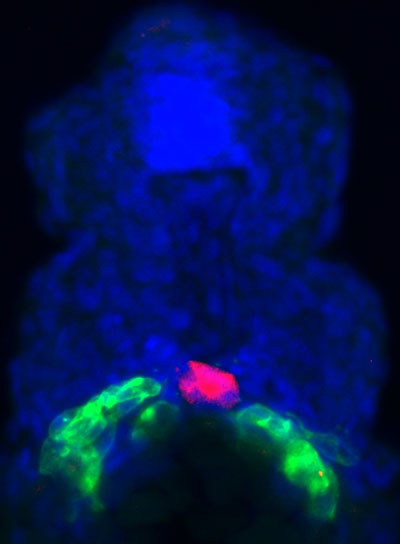
Figure: The two germ-cell clusters (green) are separated by the developing gut (red) that functions as a physical barrier.
We use the developing zebrafish embryo as a model for studying organ development, as embryogenesis in this organism occurs outside the body of the female and the embryo is translucent, allowing one to follow organogenesis at a high resolution. By monitoring the arrival of the progenitor cells in the region where the organ develops, determining the presence of proteins that could act in maintaining the cells at this site and finding out the position of other structures within the embryo that could affect the position and shape of the organ, we tried to derive general conclusions concerning the buildup of organs. The organ we use for this purpose is the gonad, a structure composed of somatic cells that support the proliferation and differentiation of germ cells, the progenitors of the sex cells, sperm and egg.
By expressing fluorescent proteins in the germ cells we were able to follow their position and movement within live developing embryos. This analysis revealed that after arriving at the site where the organ develops the cells actively migrate within this region, but do not leave it. Following the interaction of the cells with neighboring tissues revealed that the motile cells bounce off barriers that thereby limit their movement in certain directions. In addition, some nearby tissues emit signals that repel the cells and do not allow them to come near to them. Last, the motile cells progressively increase their adhesion with one another further inhibiting their motility. Together, these three activities maintain the position of the cells at a specific location in the embryo where further steps in organ development occur.
Reference
Repulsive cues combined with physical barriers and cell–cell adhesion determine progenitor cell positioning during organogenesis. Paksa, A., Bandemer, J., Hoeckendorf, B., Razin N., Tarbashevich, K., Minina, S., Meyen, D., Biundo, A., Leidel, S.A., Peyrieras, N., Gov, N.S., Keller, P.J., Raz, E. (2016). Nature Communications 7:11288. doi:10.1038/ncomms11288