


Tissue development, homeostasis, and wound healing involve cells moving collectively as groups. Collectively migrating cells use their autonomous migration machinery for locomotion, while they are connected to their neighbors through adhesive cell-cell contacts. A mechanical connection between cells has been known to be required for cells to coordinate their movements with each other. Nevertheless, an outstanding remaining question in the field was how cell-cell junctions, assembled from symmetrically organized structural components, support a signal transduction mechanism by which neighboring cells control each other’s front-back polarity.
Using monolayers of cultured human endothelial cells (HUVEC) as a model system for collective cell migration, Hayer et al. discovered that individual cells are oriented by VE-cadherin-rich membrane protrusions, “cadherin fingers”, which leader cells extend from their rear, and follower cells engulf in their front. They demonstrated the existence of cadherin fingers using two independent approaches, super-resolution microscopy and scanning electron microscopy.
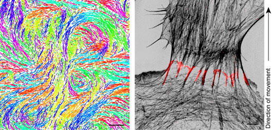
Figure - Left: Trajectories of endothelial cells migrating in a monolayer, color-coded by the direction of movement. Streams and swirls in the pattern of trajectories indicate that cells coordinate their movements with each other. Scale bar, 250 µm. Right: Cadherin fingers are asymmetric junctional structures between collectively migrating endothelial cells. 3D structured illumination microscopy (3D-SIM) of human umbilical vein endothelial cells (HUVEC) expressing the junctional marker VE-cadherin-mEGFP (red) and stained with phalloidin (greyscale). Scale bar, 2 µm.
Both approaches independently showed that cadherin fingers extending from one cell’s rear can be engulfed by the cell behind, exposing a convex or positively curved membrane surface to the cytoplasm of the follower cell. Since the type of membrane curvature of cadherin fingers exposed to the leader cell (negatively curved) is opposite from the one exposed to the follower cell (positively curved), they represent a polarized structure with respect to the direction of migration, having the potential for inducing or reinforcing polarity in the leader cell, the follower cell, or both. The authors found that the distinct types of membrane curvature were capable of selectively recruiting curvature-sensing BAR domain proteins, providing a mechanism for how membrane curvature could be translated into a signaling output.
Cadherin finger formation required an imbalance in actomyosin contractility between the rear of future leader and the front of future follower cells, VE-cadherin/catenin complexes, and Arp2/3-driven actin polymerization. Using chemical biology and optogenetics approaches, the authors showed that increased actomyosin contractility in the rear of a future leader cell is sufficient to initiate cadherin finger formation, consistent with serial polarization of adjacent cells during collective cell migration. Indeed, the orientation and location of cadherin fingers along the periphery of cells was predictive of cell turning, suggesting that cadherin fingers serve as guidance cues supporting coordinated cell movement during collective cell migration.
This study provides an intriguing solution to the long-standing question of how cell-cell junctions with symmetrically organized cadherin components mediate polarized signal transmission.
Reference
Engulfed cadherin fingers are polarized junctional structures between collectively migrating endothelial cells. Hayer, A., Shao, L., Chung, M., Joubert, L., Yang, H., Tsai, F., Bisaria, A., Betzig, E., and Meyer, T. (2016). Nat. Cell Biol. 18(12):1311-1323
Other References
Getting a grip on collective cell migration. Das, P. and Spatz JP (2016). Nat. Cell. Biol. (18):1265-1267.
Cell migration: Let your fingers do the walking. Strzyz, P. (2016). Nat. Rev. Mol. Cell Biol. Published online 30 Nov 2016; doi:10.1038/nrm.2016.160