


Recent advances in microfluidics and microscopy allowed the development of various organ- and animal-on-a-chip model systems. Now a team of researchers funded by HFSP, led by Dr. Assaf Vardi at the Weizmann Institute and Prof. Roman Stocker at MIT (now at ETH Zurich), have harnessed these technologies to develop coral-on-a-chip, the first system to allow real time microscopic imaging of reef-building corals.
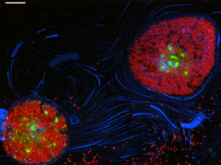
Figure: A new perspective on reef-building corals. The Coral on a Chip microfluidic platform allows the study of microscale interactions between reef corals, their environment and the diverse microorganims living in and around them. Here, two micropropagates of the reef-building coral Pocillopora damicornis, settled within a microfluidic channel, are imaged using the coral's native GFP (green) and the autofluoressence of its algal symbionts (red). Blue streaks trace the motion of microscopic particles driven by beating cilia on the coral's surface.
Coral reefs are declining at alarming rates, with some estimates predicting the loss of as many as 30% of the ocean’s coral reefs within the next 30 years. Such losses will have worldwide environmental repercussions far beyond the immediate damage to the reef ecosystem. Environmental services provided by coral reefs, such as tourism, shoreline protection and fisheries, are valued at over $30bn/year. Moreover, the stony corals that make up the backbone of the world’s coral reefs sequester 70–90 million tons of carbon each year, offsetting some of the CO2 released to the atmosphere by the burning of fossil fuels.
Much of this decilne is attributed to human activities, including global warming, environmental polution and ocean acidification. These changes to the environment were shown to induce detrimental outcomes such as coral bleaching, coral disease, and loss of fertility. Nevertheless, a mechanistic understanding of the physiological processes underlying these responses is largely missing. This is in part due to the complex interactions between corals, their symbiotic algae and the drove of other microbes living on and around them, all taking place at the realm of the microscale, defying direct observation and measurement using available tools.
This is where the HFSP-funded Vardi-Stocker collaboration steps in. The team made use of a natural stress response of hard corals, known as "polyp bail-out", to induce the expulsion of individual coral polyps from their aragonitic skeleton. They then developed a method for resettling these free polyps into pre-fabricated microfluidic channels to form the coral-on-a-chip device. Introduction of flow into the microfluidic device allows the researchers to perform live-imaging microscopy of these micropropagated corals while precisely controlling the physico-chemical conditions inside the microfluidic chamber.
In a recent publication in Nature Communications the researchers demonstrate the power of this system for the study of several processes that are at the heart of current coral research. In a first experiment, they used the Coral on a Chip system to capture the growth of individual aragonite crystals, the building blocks of the corals’ skeleton. In a second experiment, they captured bacterial pathogens as they infiltrated the oral cavity of a coral polp, revealing a hitherto little-considered route of coral infection. In a third experiment, they exposed coral micropropagates to high light intensities that induced coral bleaching on the microscope stage, capturng this globally important process for the first time at single-cell resolution.
The simple yet flexible design of the coral-on-a-chip system will enable multiple future applications. The team is already in the process of adapting this system to tracking the nutrient and carbon cycles of reef-building corals, as well as delving further into disease and bleaching processes. It is hoped that the simplicity of the system will enable its adoption by additional coral research groups around the globe, opening up new avenues for studying these fascinating and important organisms.
Reference
A coral on a chip microfluidic platform enabling live-imaging microscopy of reef-building corals. Shapiro OH., Kramarsky-Winter E., Gavish AR., Stocker R., Vardi A. (2016) Nature Communication. DOI: 10.1038/ncomms10860.