


A crucial step in exocytosis is the formation of a fusion pore that allows neurotransmitters to escape from synaptic vesicles. Although it is well established that SNARE proteins catalyze fusion, the structure and composition of fusion pores remain unknown. A major limitation in the study of fusion pores concerns their transient nature. For example, in endocrine cells, the duration of the initial open state of the fusion pore is on the order of milliseconds; the pore then either closes (kiss-and-run exocytosis) or dilates to result in full fusion. Here, we attempt to address this critical question by using the rigid framework of nanodiscs, which prevent the dilation of fusion pores.
Click on image to enlarge
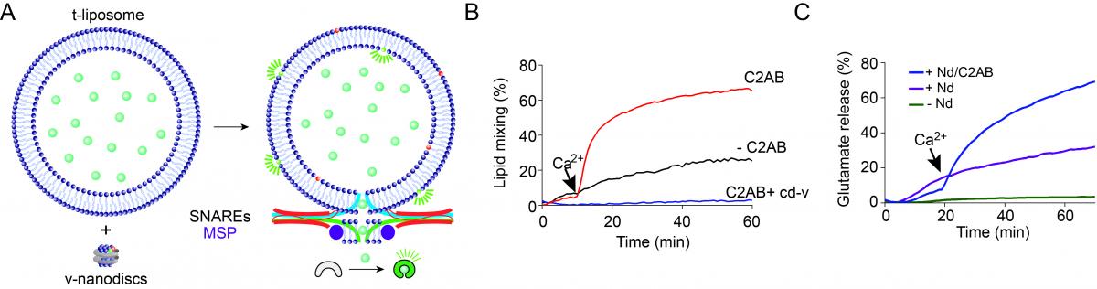
Figure: The nanodisc-SUV fusion system to study fusion pores. Fusion between nanodiscs and SUV (A) results in lipid mixing (B) and glutamate release (C), which is stimulated by Ca2+ and synaptotagmin-1.
We developed an assay in which nanodiscs bearing v-SNAREs fuse with small unilamellar vesicles (SUVs) containing t-SNAREs, in a manner accelerated by Ca2+ and synaptotagmin-1. We found that efficient Ca2+-stimulated bilayer fusion and glutamate release required only two molecules of synaptobrevin 2 (syb2) and occurred when we used 6-nm nanodiscs. In addition, we determined that the transmembrane domains of SNAREs are exposed to solvent during fusion. Collectively, these data reveal that the fusion pore is formed by a combination of lipids and SNARE transmembrane domains. We will further utilize this nanodisc-SUV fusion system to reveal the dynamics and the atomic structure of fusion pores.
Reference
Exocytotic fusion pores are composed of both lipids and proteins. Bao H, Goldschen-Ohm M, Jeggle P3, Chanda B, Edwardson JM, Chapman ER. Nat Struct Mol Biol. 2016 Jan;23(1):67-73. doi: 10.1038/nsmb.3141. Epub 2015 Dec 14.