


Our visual perception - including the ability to read this text - is dominated by the fovea. Because the fovea subtends a tiny part of the visual scene (about the size of your thumbnail at arm’s length), we make rapid eye movements to direct the fovea to key locations in a visual scene. Among mammals, the fovea is found only in humans and diurnal primates and accounts for ~50% of retinal output and ~50% of the input to the visual cortex. Despite its high spatial and chromatic sensitivity, foveal vision has a rather poor temporal sensitivity compared to peripheral vision. Past in vivo recordings of foveal output signals suggest that perceptual specializations of foveal vision originate largely in the retina itself rather than in subsequent cortical circuits. Nonetheless, we know very little about the cellular and synaptic basis of these functional specializations. This is due to a lack of intracellular recordings from foveal neurons. In this study, we make the first direct comparisons of the physiological properties of foveal and peripheral retinal neurons and the first structure-function correlation in the fovea. These experiments reveal how differences in the cellular and synaptic operation of foveal and peripheral retina can account for well-established perceptual differences in temporal sensitivity between foveal and peripheral vision.
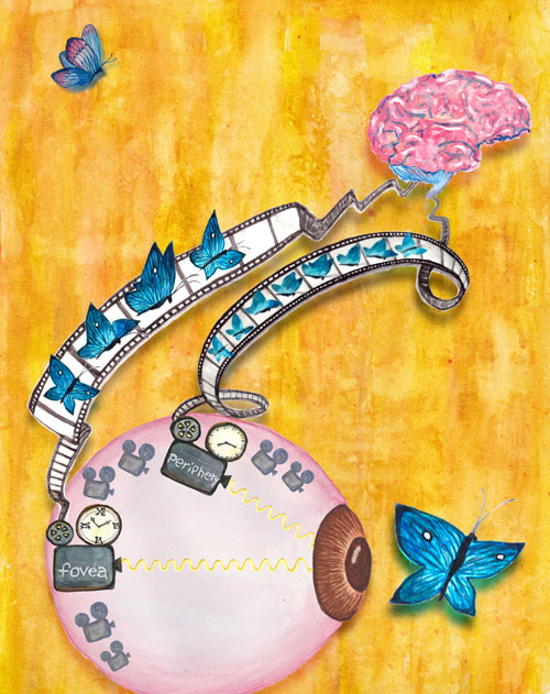
Figure: The visual information encoding the flight path of a butterfly is conveyed by cone photoreceptors depicted as old video cameras with clocks that have different gradations. The “foveal camera clock” has coarser gradations and the “camera reel” carrying the information of the butterfly’s flight to the brain with the highest spatial resolution has a slower frame rate with half as many frames compared to the peripheral camera reel. Concept by Jacob Baudin and artwork by Xiaoyun Wu.
A major theme of recent retinal work has been the surprising computational specializations of non-foveal retinal circuits, eg. direction selectivity, selectivity for complex stimulus features, etc (Ref. 1). These computations rely on inhibitory signals generated by diverse populations of inhibitory retinal neurons called amacrine cells. Surprisingly, we find that inner retinal synaptic inhibition minimally shapes responses of foveal midget ganglion cells (the dominant output neurons of the fovea). In contrast, synaptic inhibition strongly shapes responses of peripheral midget ganglion cells. Correspondingly, foveal midget ganglion cells expressed fewer inhibitory receptors on their dendrites compared to their peripheral counterparts. This lack of inhibitory input is very surprising given the importance of balanced excitation and inhibition for the operation of most sensory and cortical circuits (Ref. 2). This difference in synaptic integration between foveal and peripheral midget ganglion cells is a key insight to foveal-peripheral computational differences, but interestingly does not account for differences in temporal sensitivity between the two retinal regions.
Determining the cellular origin of human perception is an important, but rarely realized, goal in neuroscience. Our results provide a simple explanation for the perceptual observation that foveal vision is less sensitive to rapidly varying light inputs than peripheral vision. This difference in temporal sensitivity shapes our everyday visual experience, for example, it determines the frame rates of movies and refresh rates of computer monitors. To our great surprise, we found that the responses of the cone photoreceptors - the first neurons of the visual system - themselves exhibit a 2-fold difference in response time course between foveal and peripheral retina. This is nearly identical to the difference in perceptual sensitivity between the two retinal regions. Models of visual function often assume that differences between foveal and peripheral vision originate in the neural circuits that read out cone photoreceptor signals. The heterogeneity we find in the cone responses requires a reevaluation of these models and the conclusions drawn from them.
The results clearly illustrate that the fovea is not only a special structure but also a place that employs different computational strategies compared to peripheral retina. This distinction will be an important consideration in how computations are partitioned between retina and cortex. The results are also important since there is a great deal of effort to restore foveal vision in humans and our understanding of the computational structure of the fovea was largely missing.
Reference
Cellular and Circuit Mechanisms Shaping the Perceptual Properties of the Primate Fovea. Sinha R*, Hoon M*, Baudin J, Okawa H, Wong RO, Rieke F*. Cell. 2017 Jan 26; 168(3): 413-426. (*co-correspondence)
Other references
[1] Transformation of visual signals by inhibitory interneurons in retinal circuits. Jadzinsky, P.D., and Baccus, S.A. Annu Rev Neurosci. 2013, 36, 403-428.
[2]Yo How inhibition shapes cortical activity. Isaacson, J.S., and Scanziani, M. Neuron. 2011, 72, 231-243.