


One of the most intuitive strategies to classify objects in the world is to determine their color - just think of the red apple in the tree that can be spotted from a distance. This process starts in the retina, a layer behind the eye that uses a complex neuronal circuitry to transform incoming light into a neuronal code. In humans and old world primates, color processing starts by the relative absorption of light sampled by three different cone types, each absorbing preferentially in the red, green and blue range of the visual spectrum. This “RGB” system is then relayed to the brain via so-called color-opponent channels, neurons that are excited by light of one color and inhibited by light of a different one. Not surprisingly, this opponent process has been described as being an optimal strategy for relaying color information from the eye to the brain. In mammals, the “RGB” trichromacy is more of an exception than a rule. Generally, they have only two types of cones, allowing them to perceive the world in a two-dimensional color space. A more striking difference across mammals becomes evident when comparing the trichromatic system in humans with the one of many crepuscular animals that are defined as being active during twilight. Whereas the three cone types in the human retina have a “salt and pepper” distribution, the two cone types in crepuscular mammals are spatially segregated. Mice, for example, have ultraviolet and green sensitive cones, directed to the upper visual field or to the surrounding ground, respectively. Although it has been shown that mice can discriminate color, this spatial cone segregation presents a puzzle, since local comparison of the two color sensors is not possible. Therefore, it has been proposed that mice might use completely different and therefore less efficient strategies to encode color. Surprisingly, as we show in this work, the mouse retina has a dedicated cell that transmits color-opponent responses from the eye to the brain, just as expected for humans. These cells use a refined circuitry to circumvent the spatial segregation of cones, violating prior textbook expectations.
(click on image to enlarge)
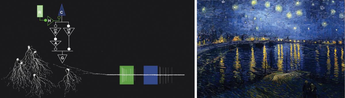
Figure: Left: Illustration of the color-opponent retinal output cells in the mouse retina, including the circuit model underlying their response properties, and an exemplified neuronal response to flashes of different color emerging from their axonal projections. Ganglion cell (G), involving rods (R), cones (C), horizontal cells (H), bipolar cells (B), and amacrine cells (A). Open/closed circles denote inhibitory/excitatory synapses. Right: Although night scenes do not have more blue in their spectra, our perception changes as exemplified by Vincent van Gogh’s ‘Starry Night Over the Rhone’.
In this work we describe a genetically defined type of output cell in the mouse retina, the so-called “J” retinal ganglion cell, which has color-opponent responses. It is activated by a decrease of ultraviolet (UV) light (OFF response) and to an increase of green light (ON response). The UV-OFF response arises in the receptive field center and is driven almost entirely by the ultraviolet cone pigment, whereas the Green-ON response derives from the surround. Although the mouse retina contains green-sensitive cones, the ON response instead originates in rods. Rod photoreceptors are known to be functional in dim lightning conditions, but are also active together with cones across a wide range of luminance, including the crepuscular light levels. Textbook knowledge professes that the rod system hijacks the existing cone pathways to transmit their information, making a comparison between cones and rods not possible. We found that this premise does not hold. We describe a neuronal circuit that compares the signals of cones antagonistically to the one of rods that processes color information in two stages. First, the center response is implemented by a cone-selective system that avoids the classical rod pathways. Second, the surround is processed through lateral inhibition of cones, unexpectedly via horizontal cells. The inhibitory horizontal cell connects to cones at the dendritic tree and to rods at the terminal arborization of its axon. Previously, it was thought that signals couldn’t propagate between these compartments. However, as we show here, it is exactly this connection that enables rod-cone opponency (see figure). Interestingly, this pathway happens to be highly conserved across mammals. In the human retina, e.g., these horizontal cells inhibit primarily the red and green cones. Under crepuscular light level, when rods and cones are active, rod signals would therefore enhance a blue percept by inhibiting red and green cones. This ‘blue shift’ experience in twilight, also known as the Purkinje effect, is probably the reason why painters add more blue to paint night scenes. Intriguingly, and in line with this phenomenon, a drug that as an off target effect enhances rod activity, namely Viagra, is known to have a peculiar side effect in some patients – a blue tint in the color percept.
To explore what ecological benefits mice might draw from UV-green color vision, we searched for objects in the natural world that would stand out in this spectrally opponent channel. Certain seeds and urine marks stand out when processed by a spectrally opponent system like the one described. Beyond its obvious excretory role, urine serves an important function for social communication among mice. Males mark their territories by squirting urine in characteristic patterns; these marks are sampled and counter-tagged by other individuals, and communicate information regarding social status. Interpretation of the tag requires physical contact because the relevant pheromones are non-volatile. Mice use primarily visual cues to recognize their territory boundaries. On that background, we propose that mice in the wild can identify urine tags visually, using the UV-green opponent color channel of the retina, which assists in approaching the tag.
Taken together, our finding changes the notion that color vision relies only on cones. In the light range where rods and cones interact, rods do contribute to the color percept in mice, as well as in humans. We expect that the discovery of this genetically-defined color-opponent channel will enable targeted studies of color processing in the brain, helping to decipher the neuronal complexity underlying the process of “seeing”.
Reference
A neuronal circuit for colour vision based on rod–cone opponency. Maximilian Joesch & Markus Meister. Nature (2016) DOI: 10.1038/nature17158.