


Executing an apparently simple task, such as grabbing a glass, involves coordinated planning from multiple brain regions. But how does that work, precisely? Intense research efforts are aimed at accurately linking brain activity to behaviour. This is particularly true for neuroscientists developing brain machine interfaces (BMIs): devices that can read neural activity and translate it into commands to an output device. Using BMIs, paralysed patients are capable of regaining control of a robotic arm.
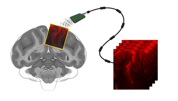
Figure: An international team of scientists transformed vascular ultrasound images from non-human primate brains into predictions about intended movements. Graphic credit: Sumner Norman.
Current BMI technologies are divided into invasive methods that rely on implantable electrodes, and non-invasive methods that suffer from a relatively low resolution. In a recent article, HFSP awardees David Maresca and Mikhail Shapiro, together with colleagues at Caltech and INSERM, show that it should be possible to combine the best of both worlds. They used functional ultrasound imaging (fUS) to record brain activity in the posterior parietal cortex of non-human primates, a brain region involved in motor planning. Neural activity corresponding to motor planning triggers tiny changes in cerebral blood flow that fUS could capture with 10 times more sensitivity than functional MRI.
Next, the team trained an algorithm to decode these fUS signals in order to find out what the primates’ intentions were. The algorithm predicted the type of behaviour the non-human primates were going to carry out (eye movement or reach), the direction of the movement (left or right), and when they planned to make the movement - all within a few seconds. Translating these findings into human neuroimaging and new, minimally invasive brain machine interfaces is an important direction of future research.
HFSP’s funding was key as it supported David Maresca (Cross-Disciplinary Fellow) who established functional ultrasound imaging capabilities at Caltech.
Funding sources
Funding was provided by a Della Martin Postdoctoral Fellowship, a Human Frontier Science Program Cross-Disciplinary Fellowship, the UCLA–Caltech Medical Science Training Program, the National Institutes of Health BRAIN Initiative, the Tianqiao and Chrissy Chen Brain–Machine Interface Center, the Boswell Foundation, and the Heritage Medical Research Institute.