


Microtubules occupy an important role in structuring living cells. They serve as intracellular transport rails and as cables that pull on chromosomes during cell division, thus ensuring the correct distribution of the genome between the daughter cells. Microtubules are extremely dynamic polymeric structures that are constantly assembling and disassembling. This process, which is called dynamic instability, is crucial to adapt the microtubule network architecture to the shape of the cell allowing the microtubules to produce mechanical forces.
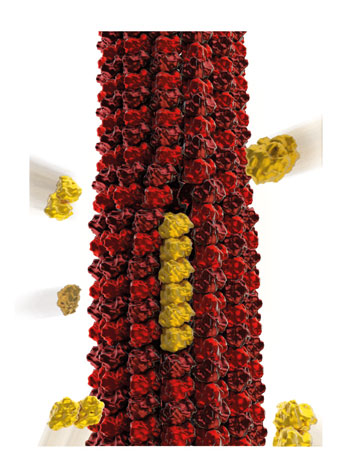
Illustration by www.illuscientia.com
Surprisingly, the dynamics of microtubules is not limited to the extremities: tubulin dimers, i.e. the building bricks of microtubules, can integrate and leave the lattice along the shaft. However, the microtubule shaft is a highly regular structure, composed of 13 so called protofilaments, which assemble in a side-by-side fashion into a hollow tube.
What mechanism enables tubulin dynamics without compromising the overall integrity of the microtubule?
Electron microscopic observations have identified small structural imperfections within the shaft. For example, the microtubule can occasionally switch between a structure composed of 13 protofilaments to a structure composed of 12 or 14 protofilaments. Analysis of the lattice dynamics using fluorescence methods and model calculations have now shown that these defects are the source of spontaneous and continuous lattice dynamics, leading to the loss or incorporation of dimers. Interestingly, the presence of imperfections confers new dynamic features to the microtubule in a trade-off resulting in a small loss in mechanical stability.
These new dynamics are intrinsic to the microtubule structure and sufficiently sensitive to be powered by thermal fluctuations only. Therefore, any microtubule binding protein may amplify or dampen it as it interacts with the lattice. These considerations open new ways to think about the regulation of microtubule stability and the related physiological processes in cells.
This animation illustrates how structural defects, resulting from imperfect microtubule self-assembly, can promote the addition/removal of new tubulin dimers (in red-orange) along the length of a microtubule, ie at a distance from the tip where the addition was believed to be restricted.