


Leukaemia is cancer of the blood and it has one of the highest cancer mortality rates. This is partly because there is a high relapse rate, as some cancer cells can survive the initial treatment. These surviving cells are often resistant to treatment, allowing the cancer to spread and become fatal. How these treatment-resistant cells survive initial chemotherapy is not well understood. One popular theory has been that they sit hiding in specific niches within the bone marrow that usually harbour blood stem cells – basic cells that can become all other blood cells.
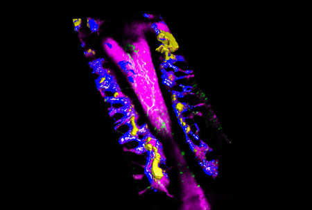
Figure: High-resolution render of unique environments in the bone marrow (blue, purple and green) as they are invaded and populated by leukaemia cells (yellow).
However, new research in mice, and validated with human samples, has revealed that certain leukaemia cells do not sit and hide. To investigate the how leukaemia works at the cellular level, the team led by researchers from Imperial College London used a technique called intravital microscopy that allows high-resolution fast imaging of live animals. The team used mice with a particularly deadly type of leukaemia called T cell acute leukaemia.
After treatment, the leukaemia cells that survived were seen moving faster than those before treatment. The researchers suggest that the act of moving itself may help the cells to survive, possibly through short-lived interactions with an array of our own cells.
The team’s investigation into leukaemia cells’ behaviour also revealed that they actively attack bone cells, which are known to support healthy blood production. The researchers believe this insight could help scientists to develop treatments to safeguard production of healthy blood cells in leukaemia patients.
HFSP funding was critical to enable this project: it contributed to the salary of multiple authors of the manuscript, covered the majority of the charges from the imaging facility we relied on during approximately the first half of the project, and animal and consumables costs. This allowed generating reliable protocols for systematic tile scanning of large bone marrow areas, followed by time lapse analysis of areas containing leukaemia cells, leading to the initial observations that T-ALL cells are continuously motile throughout the bone marrow space.
Text by Delfim Duarte and Cristina Lo Celso
Reference
T cell acute leukaemia exhibits dynamic interactions with bone marrow microenvironments. Edwin D Hawkins*, Delfim Duarte*, Olufolake Akinduro, Reema A Khorshed, Diana Passaro, Malgorzata Nowicka, Lenny Straszkowski, Mark K Scott, Steve Rothery, Nicola Ruivo, Katie Foster, Michaela Waibel, Ricky W Johnstone, Simon J Harrison, David A Westerman, Hang Quach, John Gribben, Mark D Robinson, Louise E Purton, Dominique Bonnet, Cristina Lo Celso. Nature 2016, doi:10.1038/nature19801.
Link to press release from Imperial College London