


Advanced microscopies at super-resolution or using a lattice light sheet have pushed the limits of observation of cellular events at the nanoscale. However, they and all other microscopy methods rely on traditional fluorophores or fluorescent proteins with limited optical stability, which in turn limits the long-term bioimaging of cellular events. Quantum dots (QDs) are the fluorescent nanoparticles that offer a range of optical tunability and stability superior to fluorescent proteins and probes. Yet their applications in cell biology have been limited by poor control over stoichiometry and stability of ligand display. "Direct coupling the quantum dots with endocytic ligands to facilitate their binding to cells alters the properties of ligands and thereby perturb their endocytic uptake and subsequent cellular trafficking properties," quotes Ludger Johannes, an HFSP grantee along with Yamuna Krishnan.
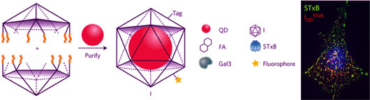
Figure: Icosahedral DNA nanocapsules, build from two halves. They host a nanoparticle such as a quantum dot (QD) and display a single biological ligand on the surface. These monofunctionalized DNA-QD-ligand conjugates can then be used to study endocytic pathways in cells adapted by different ligands such as STxB (right).
To make QDs compatible for cell biological applications, three teams — Ludger Johannes (Institute Curie) and Benoit Dubertret (ESPCI), both in Paris (France), and Yamuna Krishnan at the University of Chicago (USA) — jointly developed a new class of imaging agent that consists of:
a. A stabilized QD fine-tuned for encapsulation and with specific emission properties.
b. A DNA-based nanocage within which a QD can be encapsulated.
c. A map of this nanocapsule at single nucleotide resolution identifies locations at which an endocytic ligand can be displayed.
This results in a new class of endocytic ligand reagents that show native intracellular trafficking properties.
This was spearheaded by HFSP postdoctoral fellow Dhiraj Bhatia (holding a PhD from Yamuna Krishnan’s laboratory formerly at NCBS, Bangalore). “The advantage that these DNA devices offer is unprecedented control over the surface functionalization and robustness of coupling to diverse endocytic ligands with a strictly maintained stoichiometry of functionalization,” exclaims the young researcher.
“This platform technology now enables us to deploy bespoke endocytic probes along with advanced imaging tools like the recently acquired lattice light sheet microscopy. We can quantitatively image single molecules of proteins entering the cells over long durations of time. We are excited because this is generalizable to different kinds of nanoparticles encapsulated in these DNA nanocapsules and could lead to a new class of reagents - a powerful chemical lens to visualize unseen cell biology in membrane trafficking and biomedical research,” conclude Ludger Johannes and Yamuna Krishnan.
Reference
Quantum dot-loaded monofunctionalized DNA icosahedra for single-particle tracking of endocytic pathways. Dhiraj Bhatia, Senthil Arumugam, Michel Nasilowski, Himanshu Joshi, Christian Wunder, Valérie Chambon, Ved Prakash, Chloé Grazon, Brice Nadal, Prabal K Maiti, Ludger Johannes*, Benoit Dubertret*, Yamuna Krishnan* (* corresponding authors). Nature Nanotechnology 11 (12), 1112-1119, 2016.