


Communication between distributed divisions of the brain is facilitated by synchronized rhythmic activity. Tightly coupled brain regions serving specific functions, such as visual processing or decision making, form circuits that exhibit their own rhythmic channels of communication. Similar to electrical circuits in engineering, the brain employs feedforward and feedback circuits to regulate brain activity. For neuroscientists to ultimately influence these feedforward and feedback interactions, they must understand which brain regions are connected and how they communicate. To probe these concepts, Dr. Frohlich and his team study the ferret due to the ease of studying multiple brain areas and the folded nature of the brain. The authors utilized functional magnetic resonance imaging (fMRI), a non-invasive neuroimaging method that indexes neural activity based on blood flow, in the ferret to map out the synchronized brain regions that form functional networks.
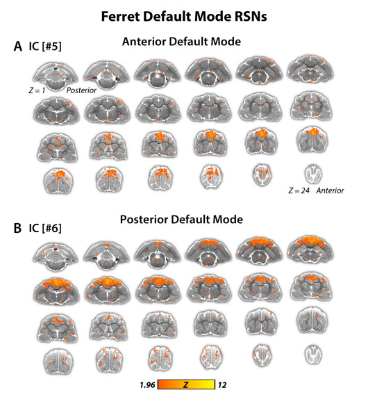
Figure: Functionally connected brain areas (highlighted in color) of the ferret default mode network
The team found functionally connected brain regions that formed sensory and motor networks, similar to what has been shown for humans and non-human primates. Importantly, the analyses revealed a higher-order brain network (the default mode network) that comprises, among other brain regions, the prefrontal cortex and the posterior parietal cortex. While these two brain regions by themselves are heavily involved in decision making and attention, the collective default mode network has been implicated in introspection, rest, and impairment in psychiatric disorders such as schizophrenia and autism.
These results provided the starting point for the manipulation of these circuits, which is the topic of an HFSP grant awarded to Drs. Frohlich and Bizley. This study is an important stepping stone in the collaboration that aims to enhance cognitive function by rational stimulation of interacting networks.
With a detailed and comprehensive map of the ferret brain, the team aims to dissect how activity in feedforward and feedback connections from the posterior parietal cortex give rise to sophisticated behavior. The ultimate goal is to selectively inactivate or rhythmically stimulate such connections in a closed-loop fashion and show changes in behavior.
Reference
Resting state network topology of the ferret brain. Zhou ZC, Salzwedel AP, Radtke-Schuller S, Li Y, Sellers KK, Gilmore JH, Shih YI, Fröhlich F, Gao W. Neuroimage. 2016 Dec;143:70-81. doi: 10.1016/j.neuroimage.2016.09.003. Epub 2016 Sep 2.