


Finding specific cells or molecular markers in deep tissue is as troublesome as finding your car at night on a full parking lot – blinking bright headlights surely help when hitting the electronic key fob. The interest in following the molecular mechanisms of health and disease and the desire to visualize the effectiveness of newly developed drugs in the complexity of an entire living organism thus faces similar challenges: available imaging modalities come with significant limitations that leave many things in the dark. Fluorescence imaging has great sensitivity and magnetic resonance imaging (MRI) provides good contrast to differentiate various tissues, but the former suffers from limited penetration depth and latter requires relatively high concentrations of reporter molecules to obtain images. Nevertheless, the visualization of small numbers of target cells or dilute molecular markers is becoming increasingly important for the concept of precision medicine to better understand the mechanisms behind different diseases or to investigate the response to newly developed drugs.
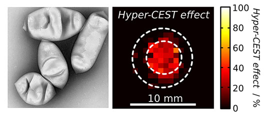
Figure: Electron microscope image of bacterial gas vesicles (left), which bind xenon reversibly as hollow protein nanostructures. Xenon MRI scan (right), in which the vesicles in the central region can be detected by a switchable contrast using the Hyper-CEST technique at an approximately 10,000-fold lower concentration than usual MR contrast media (adapted from Nature Protocols 12(10): 2050-2080 (2017) under the copyright agreement with the publisher).
The teams of physicist Leif Schröder, at the Leibniz Institute for Molecular Pharmacology, and biotechnologist Mikhail Shapiro, at the California Institute of Technology, are working on a novel approach to solve this challenge. The joint effort funded by an HFSP Program Grant investigates a new class of genetically encodable contrast agents called gas vesicles. These hollow, nano-sized protein structures are produced by certain photosynthetic microbes, where they originally evolved to function like swim bladders in fish, i.e. adjusting the floatation depth in water. These proteins were introduced by the Shapiro group as biomolecular contrast agents for ultrasound and MRI. Among their most promising applications, they combined these proteins with an advanced MRI technique originally pioneered by Schröder and colleagues, which works with the noble gas xenon known from car headlights. This technique is based on so-called laser-polarized xenon, which yields bright MRI signals already at very low concentrations. Shapiro and colleagues showed that Schröder’s technique could be used with gas vesicles because of their ability to bind the xenon.
In a joint publication in Nature Protocols, the collaborators have teamed up to improve the technique for detecting gas vesicles with laser-polarized xenon. Critically, the detection technique used by the two teams (termed Hyper-CEST) can turn the signal on and off at will through the settings of the MRI pulse sequence, such that labelled areas start blinking like headlights – just like using the key fob. This ensures reliable detection of the xenon-filled reporters. The unprecedented sensitivity allows MRI scans to be acquired in ca. 15 min for concentrations that would otherwise require millions of years when detecting the gas with conventional MRI methods. Another crucial aspect of the innovation is that these vesicles are genetically encoded, and could potentially be expressed in certain target cells that become traceable in whole organisms, a strategy that is reminiscent of the 'green fluorescent protein' (GFP), which revolutionized cell biological studies. The goal is to let the cells produce their own contrast medium and thus avoid dilution of the reporters as the cells divide: the nano-xenon headlights remain detectable from the outside.
Right now, the xenon imaging technique works only in solution. However, the collaboration is in the process of making the unconventional diagnostic procedure available for animal studies. A crucial element is the interdisciplinary approach that brings together biotechnologists, physicists, and cell biologists. The truly unconventional combination of techniques makes it also a “risky” project in the positive sense. However, most common funding agencies would be difficult to convince to support such a project. Obtaining an HFSP grant was therefore the key element to get this collaboration started. The funding provides the necessary flexibility to explore new territory. The initial results are promising and encourage the two teams in Berlin and Pasadena to move on to novel applications and to obtain fundamentally new insights.
In addition to the work on xenon MRI, the new publication describes refined protocols for imaging gas vesicles with ultrasound, with contributions from HFSP Cross-Disciplinary Fellow David Maresca, who is pursuing his postdoctoral fellowship in Shapiro's lab.
Reference
Preparation and Noninvasive Imaging of Biogenic Gas Vesicle Nanostructures. Anupama Lakshmanan, George J. Lu, Arash Farhadi, Suchita P. Nety, Martin Kunth, Audrey Lee-Gosselin, David Maresca, Raymond W. Bourdeau, Melissa Yin, Judy Yan, Christopher Witte, F. Stuart Foster, Leif Schröder, Mikhail G. Shapiro. Nature Protocols 12(10): 2050-2080 (2017).