


Even though high concentrations of the bile pigment biliverdin -or its derivative bilirubin- are an indication of a pathological condition in humans, hundreds of treefrog species in the world have physiological blood concentrations of biliverdin several times higher than those found in humans with impaired metabolic and liver function. This phenomenon is called physiological chlorosis. Remarkably, in chlorotic animals, biliverdin can stain all the tissues including the bones, a condition that has puzzled biologists for more than a century.
In our study we showed that chlorosis arose more than 40 times during the evolutionary history of anurans, spanning 11 amphibian families around the world. Using a sample of chlorotic species from different independent origins we found that biliverdin is always bound to a protein of the serpin superfamily that we termed Biliverdin Binding Serpin (BBS) (Taboada et al., 2020). We isolated, sequenced and characterized these BBSs from nine chlorotic frog species and demonstrated that all of them bind the same biliverdin isomer. We also showed that, upon binding, biliverdin changes its absorption properties giving the serpin-biliverdin complex a characteristic cyan coloration that resembles that of some components of the leaves the animals sit on.
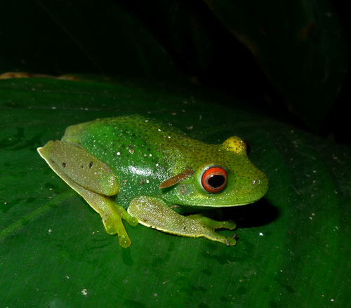
Specimen of the hylid treefrog Aplastodiscus leucopygius in Parque Natural Municipal Fazenda do Carmo, São Paulo, Brazil. Novel blue-green proteins of the serpin superfamily accumulate under the skin and modulate the color of the animals, giving them a more bluish or greenish hue in different regions of the body. Photograph: Leo Ramos Malagoli.
Serpins are a functionally diverse group of proteins, with a shared core domain and unusual structural and functional properties. They constitute the largest group of protease inhibitors in nature and are involved in biological processes such as blood coagulation, inflammation, complement activation, and angiogenesis, among many others. Remarkably, BBSs have particular amino acid substitutions that are known to preclude their normal inhibitory activity. Considering this and the fact that many of the species that have chlorosis have translucent skins with only a few yellow pigmentary cells, we explored the potential role of the novel BBS in the origin of green coloration in frogs. We quantified the concentration of BBS in the subcutaneous lymph and modeled their influence on the reflectances of the frogs’ skins. Our results demonstrated that BBS can fine tune frog coloration, explaining one underlying mechanism of crypsis in the surrounding vegetation, even in the near infrared portion of the spectrum. These results introduce a novel functional diversification of the serpin superfamily and describe an alternative way to produce green coloration in vertebrates.
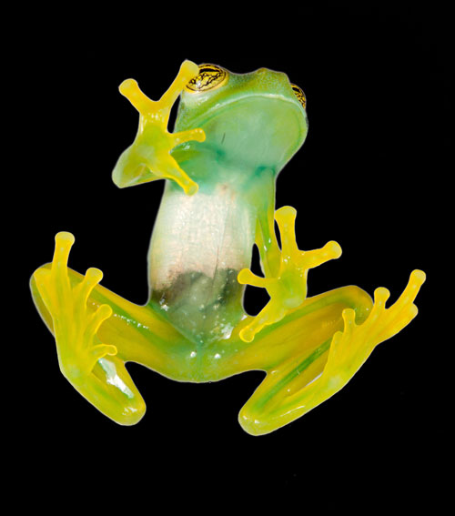
Specimen of the glassfrog Espadarana callistomma from the tropical rainforests in the Chocó region of Ecuador. Large accumulations of a novel blue-green biliverdin binding serpin can be seen under the skin of the animals and in their bones. Photograph: Santiago R. Ron.
This research started as an interdisciplinary collaboration between scientists in Argentina and Brazil, and was completed in the USA where HFSP financed the development of the pipelines to accurately sequence BBSs and the optical models. Currently, within the framework of a HFSP Long-Term Fellowship, we are studying the influence of concentration and microanatomical distribution of BBSs in the origin and maintenance of transparency and infrared reflectance in glassfrogs. We are also working in a combination of fluorescence and photoacoustic techniques to image BBSs in vivo in deep tissues.
Press coverage
How a blue protein turns tree frogs bright green (Science)