


The bacterium Pseudomonas aeruginosa is an opportunistic pathogen, responsible for the majority of hospital-acquired infections. It is highly resistant to antibiotics, rendering its treatment particularly challenging. This resistance is notably due to its capacity to form thick, disinfectant-resistant biofilms.
Bacterial cells are covered by hair-like surface appendages that allows them to move, and to interact with their environment. Such an appendage, called the Tight Adherence (Tad) pilus, causes the cells to stick to each other to form so-called biofilms, which are clumps of bacterial cells that are highly resistant to antibiotic treatment.
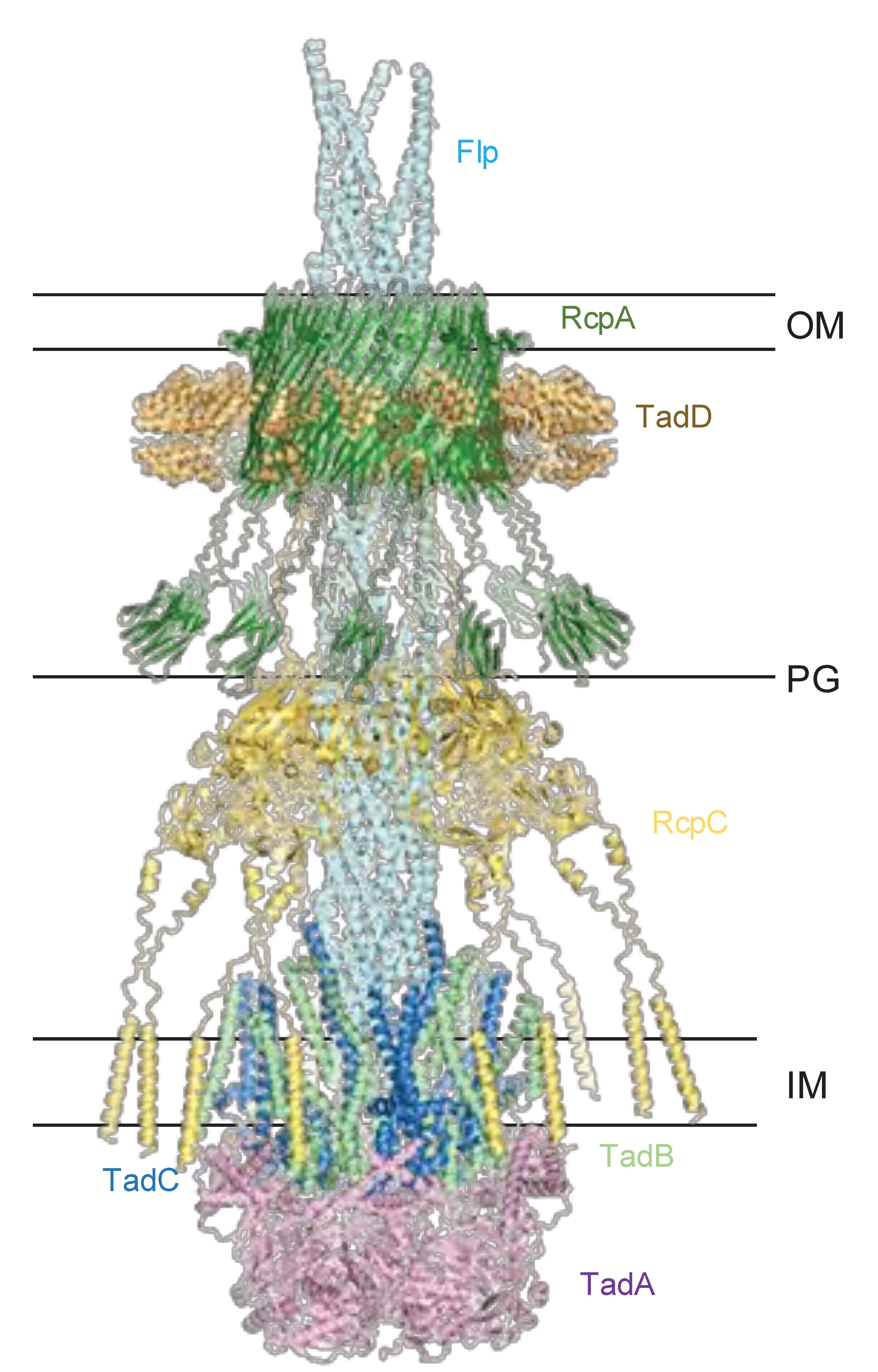
P. aeruginosa possesses Tad pili, the thin, sticky filaments on its surface that help it adhere to surfaces and form antibiotic-resistant biofilms. Tad pili are also found in many other biofilm-forming bacteria, however until now, how these pili traverse the bacterial envelope and assemble at the cell surface was not known.
HFSP Research Grant awardee Julien Bergeron and his team found that a protein called RcpC forms a ring that stretches across the cell envelope. Using high-resolution cryo-electron microscopy, they showed that this ring has a central opening just wide enough for the pilus to pass through. Furthermore, when bacteria lack RcpC, they cannot stick together or form biofilms.
This work shows that RcpC acts like a structural guide, creating a continuous tunnel that the pilus uses to exit the cell, providing unexpected molecular insights into the formation of Tad pili. These results also reveal that RcpC could be targeted by drugs to prevent biofilm formation, providing potential new avenues of treatment to combat antibiotic-resistant infections.