


When a dendritic cell, a type of white blood cell of the immune system, encounters a bacterial infection it is activated and starts to produce signal molecules. These molecules, called cytokines, are secreted to activate other immune cells which then help eliminate the infection. In this study, we investigated the cytokine interleukin-6. For secretion, vesicles containing interleukin-6 need to fuse with the cell membrane. For this process, called membrane fusion, SNARE proteins are necessary. Three or four cognate members of the SNARE protein family at both the vesicle interleukin-6-vesicles and on the cell membrane form a tight complex driving membrane fusion. SNARE proteins are highly expressed in mammalian cells and can take over each other’s function. This makes it complicated to identify the sets of SNARE proteins that secrete cytokines as well as for other intracellular membrane fusion transport routes.
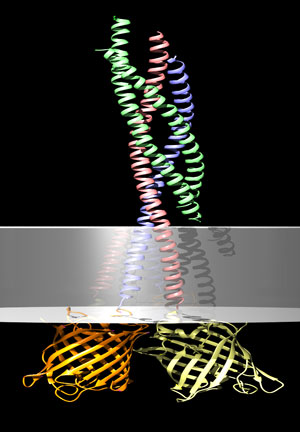
Figure. Model of the SNARE complex with conjugated fluorescent reporter proteins. The fluorescent reporter proteins are immediately juxtaposed resulting in Foerster resonance energy transfer.
In this study, we developed a novel technique based on fluorescence lifetime imaging microscopy to quantitatively visualize SNARE complex formation in live human primary dendritic cells. This enabled us to determine which SNARE proteins are responsible for the secretion of interleukin-6 by dendritic cells. We found that upon bacterial encounter, dendritic cells have increased complexing of the SNARE proteins syntaxin 4 and VAMP3. Silencing of the gene for VAMP3 reduced the amount of secreted interleukin-6.
SNARE proteins are not only essential in immune cells, but also for secretion and transport between organelles in all eukaryotic cells. Our novel technique enables to determine which of the over 30 different SNARE proteins in mammalian cells mediate these transport processes.
Reference
Fluorescence lifetime imaging microscopy reveals rerouting of SNARE trafficking driving dendritic cell activation. Verboogen, D.R.J., González Mancha, N., Ter Beest, M., & van den Bogaart, G. (2017) eLife. 6: e23525.