


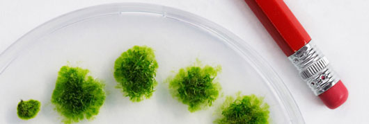
Dividing cells of plants and animals synthesize and move large amounts of membranous material to construct a division plane. How this material is routed to the division site is poorly understood. Jeroen de Keijzer and colleagues studied initial depositions of membrane during plant cytokinesis and found a striking coincidence with sites of microtubule overlaps in the phragmoplast. At the onset of cytokinesis the plant spindle is transformed into the phragmoplast that generates a membrane-enclosed template for forming a new cell wall segment. At this time, lateral contacts between antiparallel microtubules at the center of spindles shortened, very much like their counterparts in animal spindles. The team used the moss plant Physcomitrella patens to study coordination between the cytoskeleton and vesicular systems. Homologous recombination in this organism enabled the deletion of two genes encoding kinesin-4 proteins that were known for their regulatory effect on microtubule overlap length in animals. Mutant plants showed a disordered array of microtubule overlaps and thicker dividing cell wall segments that lacked structural integrity. The work thus emphasizes that zones of microtubule overlap attain a high degree of organization to precisely delimit membrane activities during plant cell division.
For the full story see the news item from Wageningen University
HFSP funding of this grant was especially helpful since it allowed the first author of the publication from the lab of Marcel Janson to work in the lab of Gohta Goshima at the Nagoya laboratory and learn the moss culturing and transformation protocols that were vital for the work conducted in Wageningen.
Reference
Shortening of Microtubule Overlap Regions Defines Membrane Delivery Sites during Plant Cytokinesis, de Keijzer J, Kieft H, Ketelaar T, Goshima G, Janson ME., Curr Biol. 2017 Feb 20;27(4):514-520. doi: 10.1016.