


































To understand how complex cognition evolves, we need to compare brain function across a wide number of species. Yet much of modern neuroscience is built on studies of only a few mammalian model systems. Birds offer a powerful opportunity to broaden this perspective. Of the roughly 11,000 bird species alive today, fewer than ten are commonly studied in neuroscience, despite their remarkable cognitive abilities. Separated from mammals by over three hundred million years of evolution and possessing very different brain structures, birds nonetheless share striking functional similarities. Notably, many species learn their songs through processes that parallel human vocal learning.
One major barrier to expanding research across species is the difficulty of measuring brain activity in non-human animals using existing technologies. To address this challenge, an HFSP-funded team led by Carlos A. Botero of the University of Texas at Austin, along with Joseph P. Culver at Washington University in St. Louis, Onur Güntürkün at Ruhr University Bochum, and Young-Gyu Yoon of KAIST, Daejeon, developed an affordable, portable, noninvasive, and easy-to-use imaging system that can be applied across a wide range of bird species. The approach builds on optical imaging methods already used in humans, which rely on light to measure brain activity. Because light can penetrate tissue and bone, changes in the reflected signal can reveal which brain regions are active.
In a recent study, led by postdoctoral fellow Kathryn C. Chenard, the researchers evaluated whether optical imaging can reliably measure brain function and connectivity in birds, using pigeons as a model system. They used widefield optical imaging to record resting-state activity across the dorsal surface of the brain, which includes regions involved in memory, such as the hippocampus, and areas important for visual processing. By analyzing patterns of correlated activity, the team was able to divide the brain into distinct functional regions that closely matched known anatomical organization. These regions were then used to construct maps of functional connectivity, uncovering how different parts of the brain interact. To further validate their findings, Chenard, Botero, and their team conducted a parallel study using MRI, a well-established method for measuring brain connectivity. The two approaches produced highly consistent results, both in identifying brain regions and in mapping the relationships between them.
In parallel with these spatial mapping efforts, the collaborative team has overcome traditional tradeoffs in high-speed imaging by developing a novel functional imaging framework based on event cameras. By leveraging an implicit neural representation algorithm to reconstruct continuous brain activity from sparse, asynchronous event streams, this technology captures rapid neuronal dynamics with exceptional temporal precision. While currently demonstrated at the scale of optical microscopy, this data-efficient approach is poised to be integrated into macroscopic diffuse optical tomography (DOT) technologies in the future, providing a powerful means to study the highly dynamic avian brain.
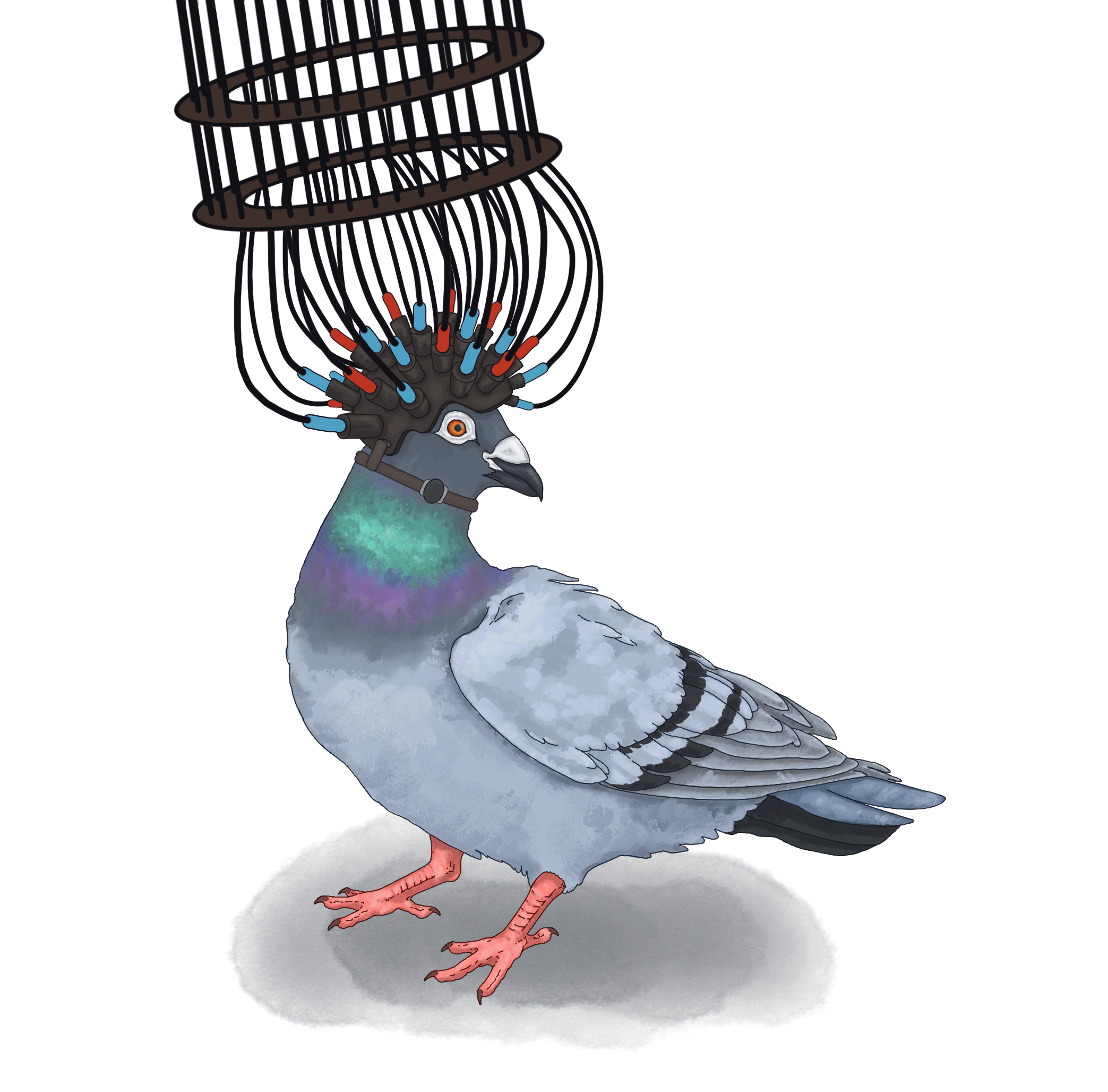
Together, these results demonstrate that optical imaging is a promising tool for studying cognition in birds. The team’s hope is that this approach will pave the way for comparative research using birds to investigate learning, memory, and spatial navigation in naturalistic contexts. Building on this work, the HFSP team is currently developing diffuse optical tomography systems capable of three-dimensional imaging across the entire avian brain. These lightweight, customizable “helmets” are currently being refined and tested in pigeons, including experiments that measure how visual brain regions respond to light stimulation. Such advances could further expand the scope of noninvasive brain imaging, facilitating comparative research across diverse species and experimental settings.