


































The bacterial flagellar motor is a rotary motor embedded in the cell surface of many bacteria. The flow of protons through a ring of motor proteins powers flagellar rotation. This rotary motor is connected to an extracellular helical filament, which, when rotated, coils to form a rotary propeller, pushing the bacterium through its environment toward more favorable locations.
Although their function remains conserved across bacteria, flagellar motors from different species have striking structural differences. While all share a common core structure, many have added additional proteins that modify the output of the motor, often to increase its torque. By focusing on the flagellar motor found in Campylobacter jejuni, the scientists supported by an HFSP Research Grant sought to characterize proteins new to Campylobacter and to probe how these additions may have occurred.
By producing very small “minicells” of Campylobacter for in situ cryogenic electron microscopy imaging and single-particle analysis of its motor, the team was able to acquire an order of magnitude more data – and of higher-quality – than ever previously obtained. Together, this fed into an electron cryo-microscopy image processing pipeline that ultimately delivered the three-dimensional structure of the Campylobacter motor to sufficient resolution for molecular interpretation. For the first time, the authors could discern the locations and orientations of almost all of the additional proteins in the Campylobacter motor.
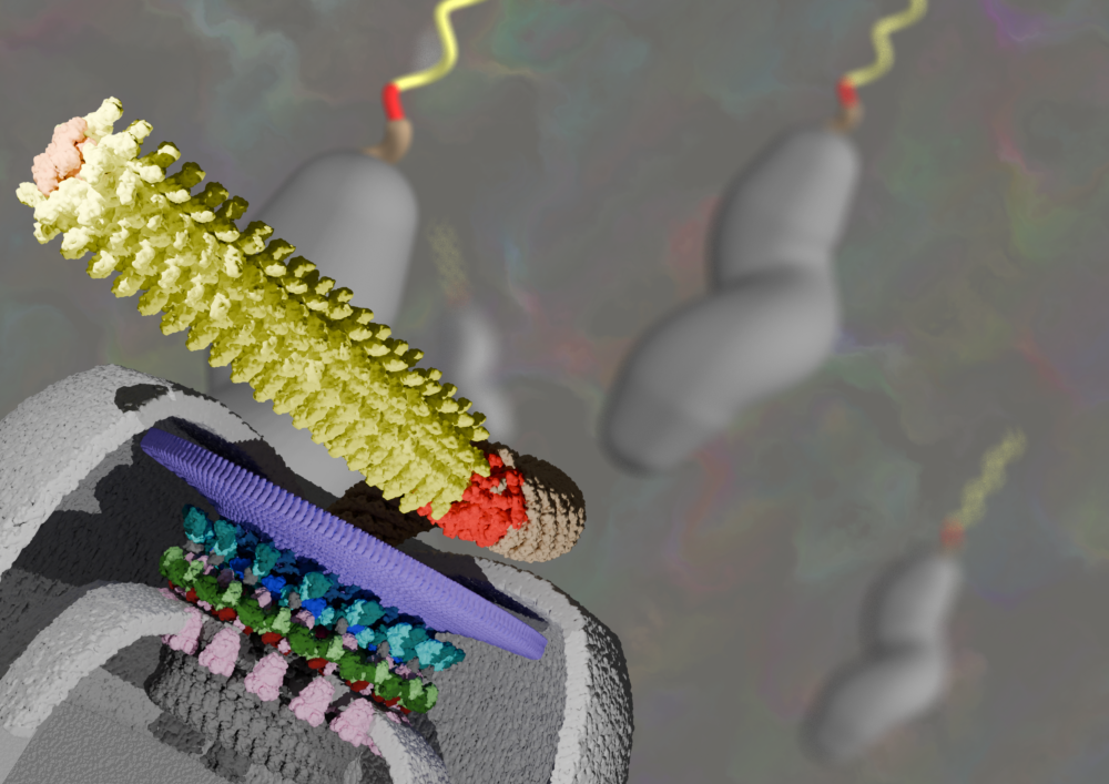
Interestingly, the structural analysis revealed similarities between newly recruited flagellar components and other proteins found in many bacteria. Most striking was the similarity of a flagellar protein, “PflC”, to a widespread family of enzymes. PflC forms a large ring-shaped lattice around the motor associated with positioning more motor proteins, resulting in increased torque. Remarkably, the enzymes that are related to PflC are found in the same compartment of the cell as flagellar motors. Furthermore, these enzymes are known to naturally coalesce to form large structures, foreshadowing the behavior of PflC in the flagellar motor. Together this suggests that the enzymes were poised to become part of the motor by already featuring characteristics that give them a selective benefit as part of an enlarged flagellar motor.
Some other features of the motor remained beyond the scope of the team’s structural determination. For example, a part of the flagellar motor of particular interest was the so-called “LP-ring”, conserved across all flagellar motors. In Campylobacter, additional proteins have been added directly onto the LP-ring, but the scientists were unable to discern the exact stoichiometry in Campylobacter jejuni. However, dynamic measurements, together with the structural data, support a model in which C. jejuni’s LP rings also comprise 26 subunits.
By building a comprehensive case-study of the flagellar motor from one species, the consortium is poised to pursue similar studies in other species for comparative work. Furthermore, by identifying the protein components that have been relatively recently recruited to the Campylobacter flagellar motor, the scientists will be able to probe the molecular mechanism of their addition and provide information on how torque is generated."